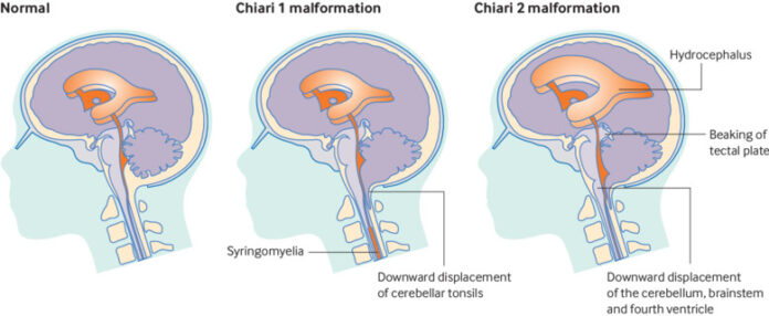
Chiari malformations (CM) occur when brain tissue extends into the upper spinal canal.
The condition is most commonly produced by congenital (existing at birth) malformations in the area of the skull wherever the brain and spinal cord join.
Type I Chiari malformations are thought to impact approximately 1 in 1,000 births in the United States. Some cases do not cause symptoms, so the actual number may be higher.
What are Chiari malformations?
The foramen magnum (pictured) is a small opening in the skull where the brain connects to the spinal cord. If the skull is misshapen then a part of the cerebellum may move through the foramen magnum.
Normally, the skull holds the brain stem and cerebellum, the part of the brain responsible for movement coordination and muscle tone. The spinal cord connects to the brain in a small opening in the skull, called the foramen magnum.
When the skull is weak or misshapen, or a drastic amount of spinal fluid is lost, parts of the cerebellum may travel down through this small opening.
If the cerebellum protrudes into the spinal canal, it can become constricted. Depending on the intensity of the pressure, problems with brain function may occur.
Circulation of the cerebrospinal fluid circulation (CSF) may also become difficult. Cerebrospinal fluid cushions the brain and spinal cord from damage. The fluid is also responsible for circulating nutrients and removing waste from the brain.
A syrinx, or fluid-filled cyst, may also form in the spinal cord, worsening the pressure.
Causes
The majority of Chiari malformations are primary or congenital cases. They are caused by physical defects in the structure of the brain, skull, and spinal column that occur during fetal development.
Conditions associated with Chiari malformations include:
- myelomeningocele, a form of spina bifida
- under-developed posterior fossa
- scoliosis
- fluid-filled cysts in the spinal cord
- malformed or thickened occipital bone
- Ehlers-Danos syndrome, a connective tissue condition
- over-crowding of the brain and spinal tissues
Most people who seek medical attention for Chiari malformations are between the ages of 20 and 40. Chiari formations are three times more common in females than males.
Recent studies indicate that there may be a genetic association with Chiari malformations.
In rare cases, severe trauma, disease, infection, or surgery can lead to the loss of large amounts of spinal fluid, causing secondary Chiari malformations.
Types
Whether primary or secondary, Chiari malformations are further classified into three different types based on the brain regions involved and the severity of the case.
The types of Chiari malformations and their most common symptoms include:
Chiari malformation type I
Type I malformations are considered the most common type of Chiari malformation. They occur when the two lower, round lobes of the cerebellum push through the foramen magnum. These lobes are called the cerebellar tonsils.
Type I malformations usually do not have any obvious symptoms. However, potential symptoms can include:
- headaches
- mobility issues
- blurry vision
- hearing loss
- insomnia
Type I malformations may go undiagnosed because the symptoms are not always present or severe, or may be associated with other conditions.
The condition is often only identified during tests for other conditions.
Chiari malformation type II
Type II Chiari malformations may cause complications such as Myelomeningocele and Hydrocephalus.
Also called Arnold-Chiari malformations, type II Chiari malformations are rarer and generally more severe than type I malformations. They occur when parts of the cerebellum and brain stem tissue have passed through the foramen magnum.
Complications from type II malformations include:
- Myelomeningocele, a form of spina bifida.
- Hydrocephalus, when the circulation of CSF fluid is blocked and collects in the head, causing it to swell. Hydrocephalus can cause death if untreated.
Most cases of type II malformations are detected during routine pregnancy ultrasounds or shortly after birth. Surgery is necessary to decrease the pressure on the brain and spinal column, and to avoid irreversible damage or death.
Chiari malformation type III
Type III Chiari malformations are extremely rare and cause debilitating, life-threatening complications.
These malformations are so extensive that the pressure causes parts of the cerebellum, brain stem, and sometimes the surrounding membranes, to burst through the skull.
Type III malformations present in very early infancy and cause more severe versions of type II symptoms. Other common complications include:
- seizures
- delays, both mental and physical
- difficulty swallowing or breathing
- a weak, muffled, or hoarse cry
- vomiting or gagging
- inability to put on weight and slow development
- excessive or uncontrollable drooling
- intense fussiness during feedings
- weak muscle tone, especially in the arms
- stiff neck
Cases are detected during routine pregnancy ultrasounds or immediately after birth. Surgery is necessary to prevent life-threatening conditions and death.
General symptoms
The range and severity of symptoms depend on the type of Chiari malformation and individual factors.
While some people never have noticeable symptoms, those who do report a similar list of problems with varying degrees of severity. A majority of symptoms become worse from sneezing and coughing.
These symptoms include:
- balance problems
- lack of coordination
- reduced fine motor skills
- neck pain
- headaches, at the back of the head
- trouble holding the head up
- vision problems, including uncontrollable eye movements (nystagmus)
- muscle weakness or numbness, especially in the upper body and extremities
- scoliosis (curvature of the spine to one side)
- dizziness
- cranial nerve compression
- hearing problems
- tinnitus, or a continuous ringing in the ears
- depression
- insomnia
- nausea and vomiting
- dulled or slowed heartbeat
Diagnosis
If a Chiari malformation is suspected, doctors will conduct a neurological exam to check for signs of cerebellar and spinal cord damage. Signs include impaired coordination, cognition, memory, sensation, and fine motor skills.
Most diagnoses require confirmation by X-rays, computed tomography (CT), or magnetic resonance imaging (MRI).
Treatment
In non-threatening cases, the first line of treatment is often routine monitoring. Once symptoms become noticeable or bothersome, surgery is usually required to prevent nervous system damage and life-threatening complications.
Multiple rounds of surgery may be required to help manage Chiari malformations.
Surgical treatment focuses on relieving pressure on the head, skull, brain tissue, and spinal cord.
The most common surgical method involves the removal of a portion of the lower back of the skull to allow more room for the tissues to expand.
Depending on the extent of the malformation, a portion of the spinal column roof and the cerebellar tonsils may be removed to increase space.
Many people require multiple rounds of surgery to manage the condition.
For infants with spina bifida, treatment focuses on closing open wounds and repositioning the spinal cord. Research shows that this option is most successful when performed in utero, or before birth.
Hydrocephalus is often treated by draining the excess fluid into another region of the body, such as the abdomen, where it can be reabsorbed.
Specialized medical professionals usually treat additional associated complications separately, including vision or coordination problems. In some instances, medications may help reduce the side effects of malformations, such as swelling.
For secondary cases, treatment of the underlying condition is often enough to help symptoms to resolve on their own.

